The Computable Plant
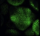
Image sequence showing cell division patterns via PIN1-GFP, in Shoot Apical Meristem (SAM),
nearby floral meristems, and the boundaries between them.
Courtesy of Marcus Heisler, Caltech. |
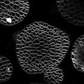
A z-stack of meristem images.
Courtesy of Marcus Heisler, Caltech. |
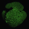
3D reconstruction of a CFP labeled flower. Animation, rotating a static 3D reconstruction and visualization of a young
arabidopsis flower using two-photon microscopy with a membrane localized CFP.
Courtesy of Marcus Heisler, Caltech. |

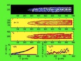
No information is available at this time.
Courtesy of The Unknown Mathematician. |
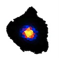
Simulation movie of the activator model simulated an a topology extracted from an experimental template.
The simulation shows the WUSCHEL concentrations. The simulation starts with zero WUS concentrations everywhere and show the
emerging WUS domain. Corresponds to Figure 7 in the paper
Jösson et al, "Modeling the organization of the WUSCHEL expression
domain in the shoot apical meristem.", Bioinformatics, 21(S1):i232-240 (2005).
Courtesy of Henrik Jönsson, Lund University. |
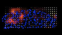
Simulation movie of the activator model simulated an a topology extracted from an experimental template.
The simulation shows the WUSCHEL concentrations. The simulation starts with zero WUS concentrations everywhere and show the
emerging WUS domain. Corresponds to Figure 6 in the paper
Jösson et al, "Modeling the organization of the WUSCHEL expression
domain in the shoot apical meristem.", Bioinformatics, 21(S1):i232-240 (2005).
Courtesy of Henrik Jönsson, Lund University. |

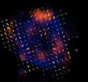
Movie of the simulation presented in Fig. 5A of the paper:
Jönsson et al, "An Auxin-driven polarized transport model for phyllotaxis," PNAS 103(5):1633-1638 (13 Jan 2006).
Shown is a simulation of the cell-based model, including cellular growth and mechanical interactions, on a shoot-like topology.
The initial auxin concentrations are random, and the simulation results in a spiral pattern.
Additional supporting information for this paper is available.
Courtesy of Henrik Jönsson, Lund University. |
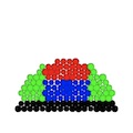
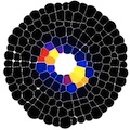
Two-dimensional geometrical simulation of four mutually repulsive self-activating proteins
representing the four morphological regions: central zone (red); peripheral zone (green);
rib meristem (blue); differentiated stem cells below the meristem (black).
Courtesy of Henrik Jönsson, Lund University. |

Three dimensional reconstruction of full meristem showing calculated nuclear centers
(red dots) overlaying CFP marked nuclei.
Courtesy of Marcus Heisler, Caltech. |

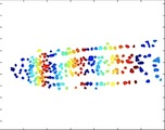
No information is available at this time.
Courtesy of Tigran Bacarian, University of California Irvine. |
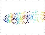
No information is available at this time.
Courtesy of Marcus Heisler, Caltech. |
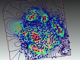
No information is available at this time.
Courtesy of Marcus Heisler, Caltech. |
No information is available at this time.
Courtesy of Tigran Bacarian, Univeristy of California, Irvine. |
No information is available at this time.
Courtesy of Tigran Bacarian, Univeristy of California, Irvine. |
No information is available at this time.
Courtesy of Tigran Bacarian, Univeristy of California, Irvine. |

No information is available at this time.
Courtesy of Eva-Maria Schötz, Laboratory of Carl-Philipp Heisenberg, Max Planck Institute of Molecular Cell Biology and Genetics, Dresden. |
No information is available at this time.
Courtesy of Marcus Heisler, Caltech. |